Global scholars gain international professional and personal experiences
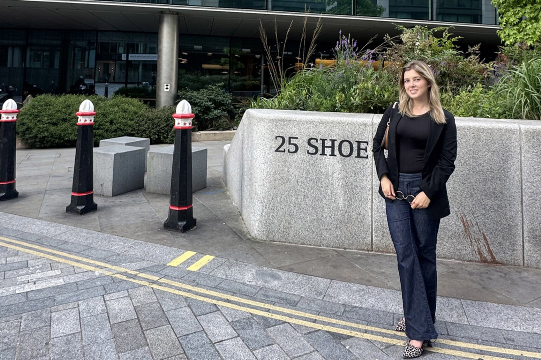
Natali Belusic wanted to learn about business while having the opportunity to be immersed in an English-language environment. RIT Croatia was the perfect choice to achieve that, and it opened the doors for international learning and experience. After landing a summer internship at Goldman Sachs in London, Belusic is now studying on RIT’s main campus in Rochester as part of the global scholars program. The global scholars program brings students from RIT’s international campuses to study for up to two terms at the Rochester campus. The experience gives students a chance to diversify their cultural perspectives, expand their academic horizons, and build their professional networks. The program, in its 15th year, welcomed its largest group of 77 students this fall. When it began in the 2010-2011 academic year, there were just six global scholars. Alumni of the program have gone on to successful careers. One former student helped build a system for the International Space Station then went on to Columbia University to pursue a master’s degree. Another is the curator of the National Gallery of Kosovo. Others work for companies including TikTok, Coca-Cola, and Amazon. Belusic is studying international business. She already gained valuable real-world experience while interning at Goldman Sachs’ risk division for nine weeks. While she didn’t think she would get the internship because of how selective and competitive it is, she felt well-prepared when applying. “RIT helped me prepare for the internship in terms of courses that I had and helped me shape my résumé and cover letter, give me knowledge on how to approach interviews, and network, as well,” she said. Now Belusic is using her time in the U.S. to broaden her academic and professional portfolio before graduating this spring. Then she will begin a full-time role back at Goldman Sachs in London next summer. “I decided to take the opportunity of additional courses and resources here that are not offered in Croatia,” said Belusic. “For example, the Bloomberg terminals that we can use and social perspective classes gave me benefits for my academic journey.” Another opportunity Belusic had while studying in the U.S. was to publish a research paper with one of her professors from Croatia. She did this through the honors program with the International Conference on Business, Management, Economics, and Information Systems hosted by The City University of New York. Experiences like Belusic’s are what make the global scholars program and RIT’s international connections a benefit for students.The program gives international students the chance to complete minors and participate in research projects that may not be offered at their home campuses. These benefits lead to impressive career opportunities. While Belusic has always been interested in working abroad, it may seem daunting for some students to travel and study in a different country. However, Belusic explained that going to a new place with a new environment helps a person learn more about themselves and how they handle different situations. “Study-abroad experiences have always exceeded my expectations,” said Belusic. “There are so many reasons to do it and so many support systems. Say yes to all the opportunities wherever you may go.”